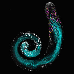
Under Pressure: Mural Cells Wrap Tightly around Blood Vessels
Under Pressure: Mural Cells Wrap Tightly around Blood Vessels
Collections: Image Award Winners, Cancer Discovery Science
2011 Award Winner
Kwabena Badu-Nkansah
Hynes Lab
Koch Institute at MIT, MIT Department of Biology
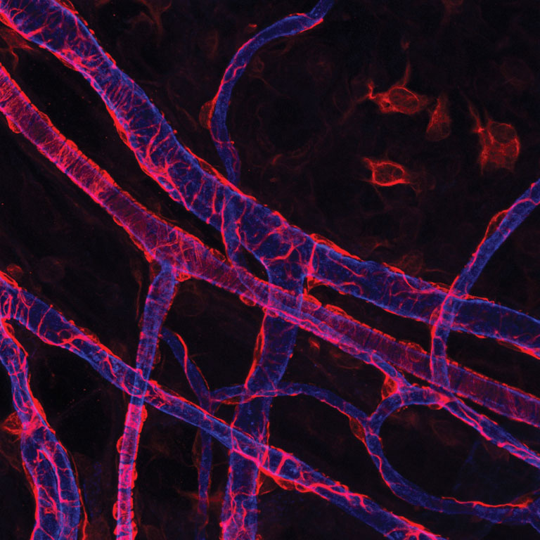
Confocal micrograph
Kwabena Badu-Nkansah works to piece together the puzzle of how blood vessels form, both in healthy cells and in cancer cells. This image reveals a small piece of the puzzle.
The veins and arteries seen here (blue) are wrapped in specialized cells called mural cells (red). From the latin murus for wall, these cells provide structural support and keep the contents of the blood vessels under pressure. Although they appear normal, these mural cells are genetically aberrant – Kwabena has engineered them to lack a certain protein implicated in cancer. By examining the formation of mural cells with and without this protein, Kwabena can better understand the role of the protein in both normal and cancerous biology.
Video
Kwabena Badu-Nkansah explains how and why he captured this image of blood vessel formation.